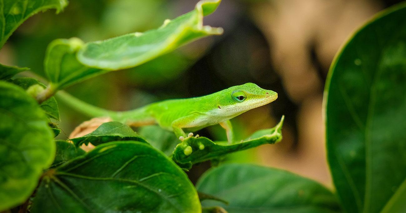
Among the amniotes, reptiles display the greatest regenerative capacity. Young alligators can regrow their tail, which includes cartilage, connective tissues, vasculature, nerves, and skin. Lizards display the remarkable ability to regenerate their tails after self-amputation, forming new muscle groups and hyaline cartilage, a tissue type that current medical therapies are unable to regenerate. The green anole lizard, Anolis carolinensis, has been selected for genomic sequencing, and this gives us a unique opportunity to examine the molecular control of regeneration. In particular, lizards are able to form new hyaline/articular cartilage, a tissue type that mammals are unable to regenerate.
Histological studies of the repair process in the A. carolinensis lizard have made clear that the regenerated tail has a quite different musculoskeletal structure from the original. A single hyaline cartilage tube that forms to replace the missing axial vertebrae, but the myoblasts are able to generate de novo myotomal blocks of tissue. In addition, the ependyma and tail nerves are also regenerated. A classic blastema-like structure does not form, and the regenerated tissues appear to derive from migrating mesenchymal cells rather than from tissue adjacent to the autotomized zone. We are currently studying this remarkable process by molecular analysis of transcriptome, microRNAome, and proteome. Comparing and applying these results to wound healing of the tail in mammals, these findings will help development of innovative therapies for regeneration and repair of cartilage and muscle.
